Microscopy
The microscopy-based methods, such as confocal microscopy, live cell imaging, immuno-electron microscopy, provide unique and indispensable information for biomedical sciences in the post-genomic era.
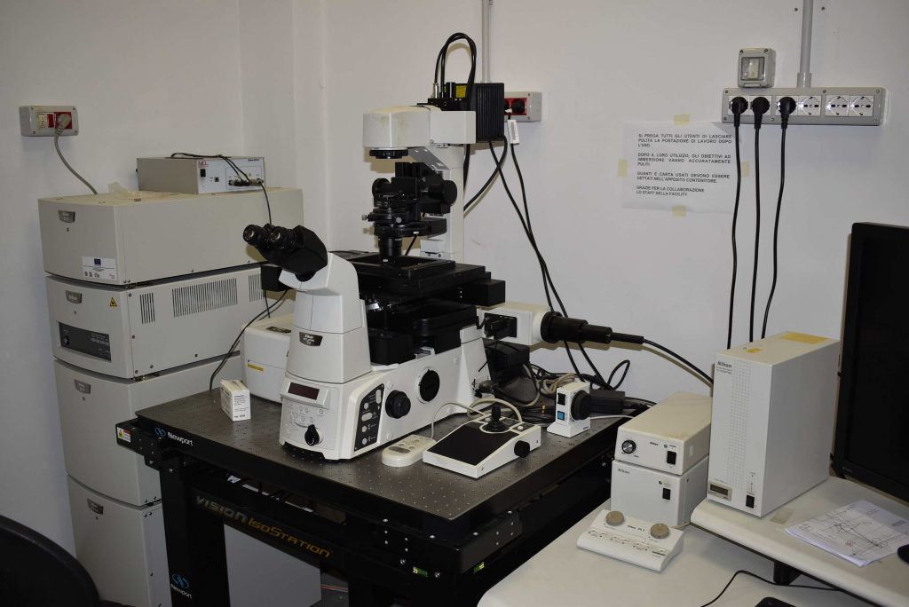
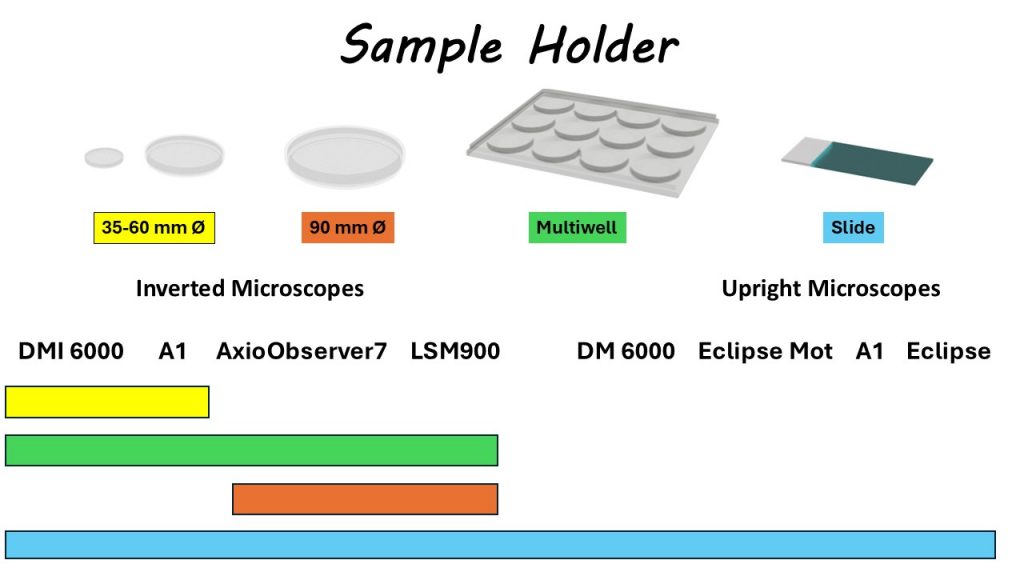
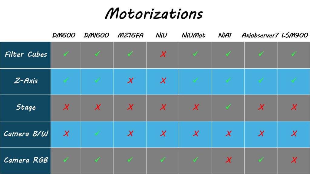
Since 2009, the IGB has established an Integrated Microscopy Facility (IGB-IM), located on the first floor of Building 3 in the Research Area of Naples 1. The IGB-IM is organized into functional areas and equipped with various instruments for sectioning and specimen preparation, as well as different types of microscopes (stereomicroscopes, upright and inverted automated microscopes for light/fluorescence microscopy, confocal microscopes, and a transmission electron microscope). In addition, workstations and softwares for image processing and analysis are available. The staff consists of one technologist and one specialized technician.
The aims of the IGB-IM Facility are to ensure the proper management and use of high-complexity equipment, to provide technical and scientific support for sample preparation, and to offer continuous guidance to all research groups in experimental design, the development of new protocols, the selection of instruments, imaging, and data analysis. The IGB-IM offers a range of techniques for the morphological, ultrastructural, and functional study of biological processes.
To this end, the IGB-IM staff organizes ongoing training for both beginners and experienced microscopy users and collaborates with other CNR Institutes, universities, and public or private companies. Theoretical and practical training courses for new users of the Facility are held throughout the year (usually in March–April and October–November) to promote optimal use of the instruments and reduce management costs. In collaboration with various companies, the staff also organizes courses on new instruments and techniques for the preparation of biological samples.
The IGB-IM Facility actively collaborates on research projects with researchers from the CNR, universities, and public or private organizations.
IGB users can access the booking page using the following link: booking service
Equipment
- Microscope
- Sample Preparation
- Useful links
UPRIGHT
INVERTED
CONFOCAL


https://www.leica-microsystems.com/products/microscope-software/p/leica-las-x-ls/downloads/

https://www.microscope.healthcare.nikon.com/it_EU/products/software/nis-elements/software-resources

https://www.zeiss.com/microscopy/en/products/software/zeiss-zen-lite.html#downloads

https://www.thermofisher.com/order/fluorescence-spectraviewer/#!/
![]()

